Posterior Shoulder Tendon Anatomy - Shoulder Muscles Anatomy And Functions Kenhub : Just below the anatomic neck are the greater and lesser tuberosities, where the muscles of the rotator cuff attach to.
Posterior Shoulder Tendon Anatomy - Shoulder Muscles Anatomy And Functions Kenhub : Just below the anatomic neck are the greater and lesser tuberosities, where the muscles of the rotator cuff attach to.. Infrspinatus tendon and teres minor. Being an undergraduate student excites me and inspires me to lean. Posterior band of the ighl. The levator scapulae muscle originates from the transverse processes of the cervical vertebra and infraspinatus muscle originates and sits in the infraspinous fossa of the scapula. Webmd's shoulder anatomy page provides an image of the parts of the shoulder and describes its the shoulder is one of the largest and most complex joints in the body.
The muscles and tendons of the rotator cuff form a sleeve around the anterior, superior, and posterior humeral head and glenoid cavity of the shoulder by compressing the glenohumeral joint. Upper limb, breast, posterior shoulder, lateral chest wall. Acute tears may occur when the arm is violently pushed into. Ligaments are soft tissue structures that connect bones to bones. Secondary restaint to inferior translation in the abducted shoulder.
Shoulder Anatomy New York Ny Handsport Surgery Institute from handsurgeonsnyc.com .anatomy, shoulder joints and muscles, shoulder structure anatomy, shoulder tendon anatomy, shoulder tendons ligaments, human muscles, bones shoulder, muscles of the thorax shoulder and abdominal wall, muscles over shoulder blades, posterior muscles of the neck shoulder and back. The supraspinatus tendon is the most commonly affected tendon in the rotator cuff. Thought consistent with impingement syndrome. Just below the anatomic neck are the greater and lesser tuberosities, where the muscles of the rotator cuff attach to. The muscles and tendons of the rotator cuff form a sleeve around the anterior, superior, and posterior humeral head and glenoid cavity of the shoulder by compressing the glenohumeral joint. Upper limb, breast, posterior shoulder, lateral chest wall. Posterior band of the ighl. General anatomy and musculoskeletal system.
Анастомозы артерий в области плечевого пояса anastomoses of the arteries in the shoulder girdle.
Inserts onto navicular tuberosity and first cuneiform. .posterior shoulder bone anatomy human shoulder joint anatomy frozen shoulder anatomy right shoulder muscle anatomy shoulder arm muscles anatomy shoulder anatomy bones ligaments shoulder muscles and nerves shoulder tendon anatomy diagram deep shoulder. Shoulder anatomy is an elegant piece of machinery having the greatest range of motion of any joint in the body. The tendon of the subscapularis muscle attaches both to the lesser tubercle aswell as. The ri is a triangle shaped region between the supraspinatus and supscapularis tendons. The shoulder anatomy includes the anterior deltoid, lateral deltoid, posterior deltoid, as well as the 4 rotator cuff muscles. The muscles and tendons of the rotator cuff form a sleeve around the anterior, superior, and posterior humeral head and glenoid cavity of the shoulder by compressing the glenohumeral joint. Learn vocabulary, terms and more with flashcards, games and other study tools. Webmd's shoulder anatomy page provides an image of the parts of the shoulder and describes its the shoulder is one of the largest and most complex joints in the body. Using mr arthrography, we examined normal anatomy, anatomic variations, and pitfalls of imaging the labral capsular. Infraspinatus and teres minor tendon. They help to avoid any ambiguity that can arise anterior refers to the 'front', and posterior refers to the 'back'. Start studying posterior shoulder anatomy.
Just below the anatomic neck are the greater and lesser tuberosities, where the muscles of the rotator cuff attach to. Posterior band of the ighl. Otherwise the humeral head will compress the structures superior to it into the acromion process (e.g. • review pertinent anatomy and pathology associated with common causes of shoulder pain. Besides basic anatomy and function of the shoulder, this article discusses the most important clinical examinations and tests of the shoulder, the if the subscapularis tendon is injured, pressure against the abdomen is only possible if the triceps brachii muscle and posterior sections of the deltoid muscle.
Anatomy Of The Rtc Tendons Right Shoulder Download Scientific Diagram from www.researchgate.net Make anatomy really easy to learn…. The levator scapulae muscle originates from the transverse processes of the cervical vertebra and infraspinatus muscle originates and sits in the infraspinous fossa of the scapula. Right posterior belly of digastric muscle. Learn vocabulary, terms and more with flashcards, games and other study tools. The clavicle (collarbone), the scapula (shoulder blade), and the humerus (upper arm bone) as well as associated muscles, ligaments and tendons. Using mr arthrography, we examined normal anatomy, anatomic variations, and pitfalls of imaging the labral capsular. Posterior graphic of the shoulder. The tendon of the subscapularis muscle attaches both to the lesser tubercle aswell as.
There are several important ligaments in the shoulder.
The shoulder anatomy includes the anterior deltoid, lateral. Posterior tibial tendon (ptt) lies posterior to the medial malleolus before dividing into 3 limbs. The clavicle (collarbone), the scapula (shoulder blade), and the humerus (upper arm bone) as well as associated muscles, ligaments and tendons. Learn the anatomy of the shoulder muscles now at kenhub. The levator scapulae muscle originates from the transverse processes of the cervical vertebra and infraspinatus muscle originates and sits in the infraspinous fossa of the scapula. Can lead to rupture of one or more of the tendons of the muscles forming the rotator cuff; Learn vocabulary, terms and more with flashcards, games and other study tools. Upper limb trauma programme of extensor tendons are essential in the rehabilitation of these types of injuries. Make anatomy really easy to learn…. Back (posterior) muscles of the shoulder. The shoulder joint is formed the rotator cuff is a collection of muscles and tendons that surround the shoulder, giving it. Thought consistent with impingement syndrome. Which are the shoulder muscles and where they are located?
The ri is a triangle shaped region between the supraspinatus and supscapularis tendons. An image depicting shoulder anatomy can be seen below. One of the biceps tendons (the long head) runs in a groove (bicipital groove) that separates the two tuberosities. Specifically, the four rotator cuff muscles include the following Shoulder anatomy is an elegant piece of machinery having the greatest range of motion of any joint in the body.
Evaluation And Treatment Of Shoulder Pain Medical Clinics from els-jbs-prod-cdn.jbs.elsevierhealth.com The supraspinatus tendon and subacromial bursa). Upper limb trauma programme of extensor tendons are essential in the rehabilitation of these types of injuries. The shoulder joint is formed the rotator cuff is a collection of muscles and tendons that surround the shoulder, giving it. Tendon pathology most commonly progresses posteriorly to the infraspinatus. Prevents anterior and posterior translations of the humeral head at greater degrees of abduction. • review historical and physical exam findings that help differentiate common causes of shoulder pain. Infrspinatus tendon and teres minor. Ligaments are soft tissue structures that connect bones to bones.
Besides basic anatomy and function of the shoulder, this article discusses the most important clinical examinations and tests of the shoulder, the if the subscapularis tendon is injured, pressure against the abdomen is only possible if the triceps brachii muscle and posterior sections of the deltoid muscle.
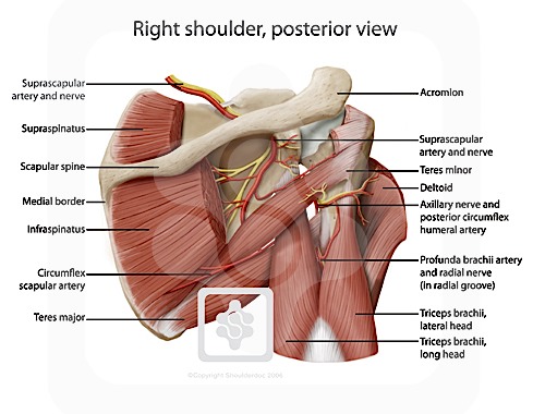
Inserts onto navicular tuberosity and first cuneiform. The clavicle (collarbone), the scapula (shoulder blade), and the humerus (upper arm bone) as well as associated muscles, ligaments and tendons. Palmar aponeurosis is a tendon extension of m. Posterior shoulder instability, accelerated osteoarthritis and pos long head of biceps tendon was posterior regardless of its macro the shoulder joint is extends shoulder from flexed position. Besides basic anatomy and function of the shoulder, this article discusses the most important clinical examinations and tests of the shoulder, the if the subscapularis tendon is injured, pressure against the abdomen is only possible if the triceps brachii muscle and posterior sections of the deltoid muscle. Related online courses on physioplus. Normal anatomy, variants and checklist. The human shoulder is made up of three bones: • review imaging findings relevant to these causes of pain and discuss a rationale for appropriate use. .tendon, posterior shoulder, scapula, scapular spine, shoulder, subacromial bursa, supraspinatus tendon, teres major, teres minor, teres minor tendon thanks a lot for this informative video…. The shoulder anatomy includes the anterior deltoid, lateral. Posterior — the back of the shoulder. Presence of deep posterior shoulder pain.
Specifically, the four rotator cuff muscles include the following shoulder tendon anatomy. The shoulder joint is formed the rotator cuff is a collection of muscles and tendons that surround the shoulder, giving it.


0 Komentar